
Vial Stopper Coring: Needle Gauge, Bevel Angle and How Rubber Ends Up in Solution
A needle through an elastomeric septum is a cutting operation, and sometimes it punches out a fragment that falls into the container — here is what the measured incidence actually is, and where the published advice contradicts itself.
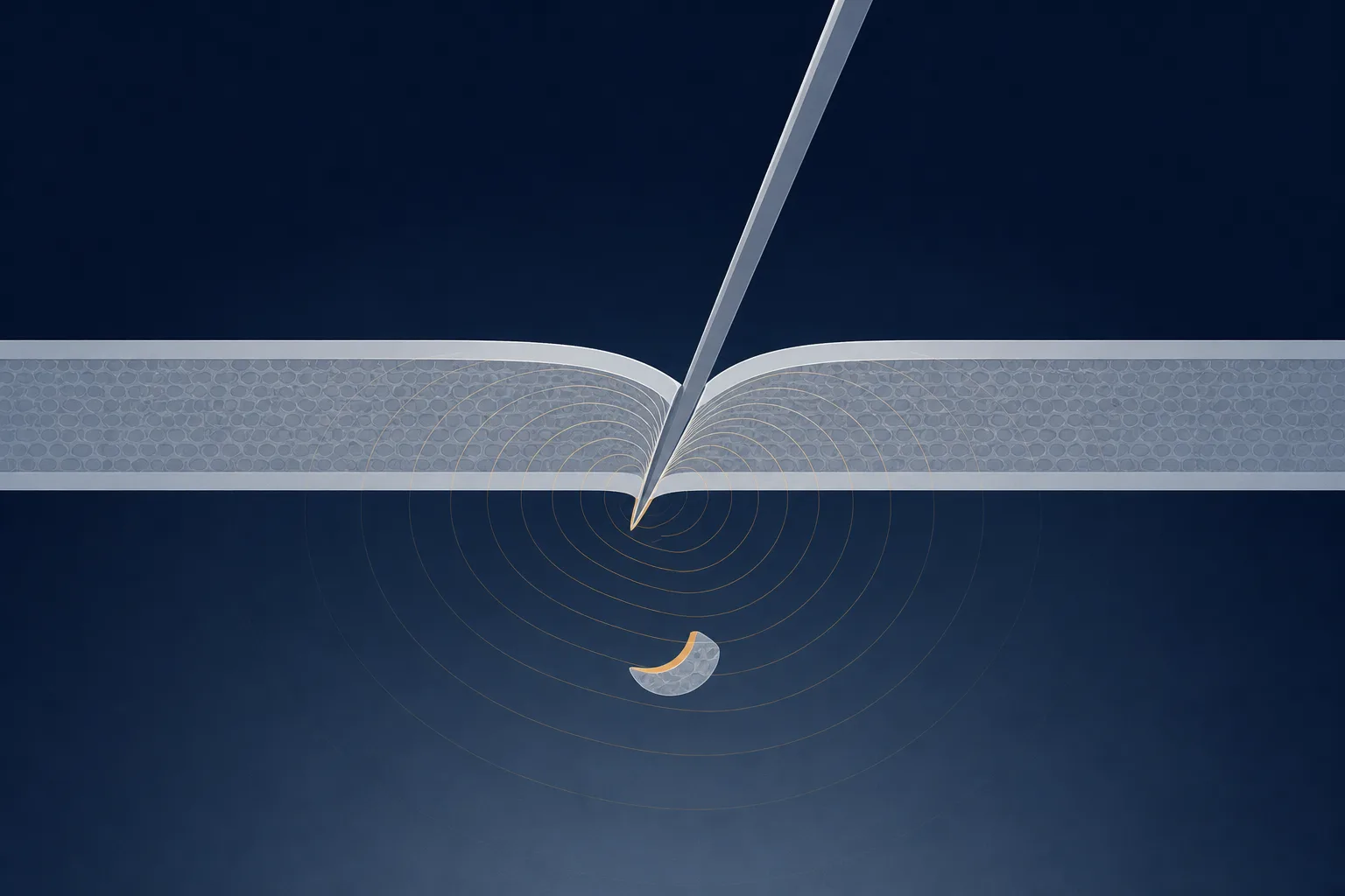
Coring happens because a needle passing through an elastomeric septum is doing cutting work, not tunnelling. The sharpened edge can complete a crescent-shaped cut through the rubber rather than parting it, and the severed piece is carried into the container behind the shaft. Three things are controllable: bevel-up entry at an oblique angle, rotated to vertical as the tip passes through [1]; a fresh needle for every entry; and the access device, where a blunt fill cannula turns out to be the highest-risk option in the published comparisons rather than the safest [2][8]. None of it reduces the risk to zero. Measured incidence spans roughly 3% to 73% of entries depending on device, closure, and how hard anyone looked [3][4].
This article treats the closure as what it is at a bench: a compendially specified elastomeric component with published test methods, penetrated by a device with a defined geometry. The compounds handled alongside it are supplied for research use only, and what follows concerns septum mechanics, the resulting particulate, and the records kept about both.
What coring is, and what a core actually looks like
A hypodermic bevel is a chisel — a leading point with two cutting edges behind it that open the material outward. When geometry, entry angle and the elastomer's resistance line up unfavourably, the leading edge completes an arc back on itself before the tip clears the septum, and the enclosed piece of rubber sits in the lumen or drops into the headspace. Fragmentation is the broader term used in the standards, covering both the intact plug and the shavings a needle produces when it scrapes rather than punches [10][13].
The fragments are not always dramatic. Rubber particles recovered during preparation of intra-articular injections averaged about 77 µm long and 36 µm wide, with individual pieces running from roughly 29 µm to 214 µm [7]. The compendial fragmentation test assumes a fragment larger than about 50 µm is visible to the unaided eye [10]. So some coring events produce something you can see against a light, and some do not.
How often coring happens: the published numbers disagree
There is no single incidence figure, and the spread is itself the finding. An observational series across routine anaesthesia practice recorded coring in 3.1% of vial accesses [3]. Investigators who collected insulin cartridges from thirty hospitalised patients and filtered the contents found rubber fragments after 73% of first entries and in 97% of cartridge remainders [4]. Same phenomenon, different scrutiny, different closures, different numbers of punctures.
| Study | Access device | Reported incidence | What was counted |
|---|---|---|---|
| Insulin cartridges from 30 patients, 2001 | 31-gauge pen needle | 73% of first entries; 97% of remainders | Fragments recovered by filtration |
| Prospective device comparison, 2014 | 18-gauge sharp vs blunt plastic | 4.2% sharp; 40.8% blunt | Visible coring per vial entered |
| Observational series, routine practice, 2015 | Mixed devices in routine use | 3.1% overall; 9.9% blunt plastic cannula | Coring per vial access |
| Bench study, multi-dose propofol vials, 2022 | 18G, 20G, 21G at 45° and 90° | 17.3% overall; 38% at 18G; 56% at 18G/45° | Coring after repeated punctures |
| Intra-articular preparation study, 2021 | 18G, 23G, and 18G with 5 µm filter | 10%, 33% and 0% | Particles in the withdrawn aliquot |
Read the fourth column before the third. Counting visible plugs and filtering the withdrawn liquid through a fine membrane are not the same measurement, and the second will always report more. The useful output of this literature is the direction of each effect, not its magnitude.
Needle gauge: the evidence points in two directions
A larger bore removes more material, and one line of evidence behaves accordingly. Puncturing multi-dose propofol stoppers with 18G, 20G and 21G needles produced coring in 17.3% of 150 samples, with 18-gauge accounting for the highest share at 38%; bore size dominated regardless of angle [6]. The 2025 joint alert from the Anesthesia Patient Safety Foundation and ECRI/ISMP advises smaller-gauge access, suggesting 21-gauge over 18-gauge where practicable [8], and a review by the American Society of Retina Specialists' safety committee lists larger gauges among the recognised risk factors, alongside perpendicular entry, multiple-dose containers and thicker stoppers [9].
The contradiction should be stated, not smoothed over. In the intra-articular study the opposite appeared: particles came from 10% of vials entered with an 18-gauge needle but 33% entered with a 23-gauge, and the authors noted explicitly that thinner needles cored more in their hands [7]. The insulin work, using a 31-gauge pen needle, produced the highest incidence in the literature [4]. One reconciliation is that fine needles core small pieces readily while wide needles core large pieces rarely, so the answer depends on whether a study counts particles or plugs — a hypothesis, not a finding.
Blunt fill needles are not the safe option they look like
Blunt plastic cannulas exist to reduce sharps injuries, and it is tempting to assume a device with no cutting edge cannot cut a stopper. The measured result is the reverse. A prospective comparison found coring in 102 of 250 vials (40.8%) entered with a blunt needle against 9 of 215 (4.2%) with a sharp 18-gauge hypodermic [2]. An independent observational series agreed, the blunt plastic cannula being the worst device at 9.9% of uses against 3.1% overall [3]. A blunt tip does not initiate a clean puncture: it stretches the septum until the elastomer tears, and a tear detaches material far more readily than an incision.
This is now formal guidance. The April 2025 alert states that while coring occurs with any needle used to access a flexible stopper, the highest risk appears to be associated with blunt needles, and recommends sharp needles unless the container's labelling specifies a blunt access device [8]. Some closures are validated for a specific blunt cannula, and there the manufacturer's instruction is the authority.
Bevel orientation and entry angle: the technique actually described
The technique cited most often comes from a 2007 note in Anesthesia & Analgesia: present the needle with the bevel opening facing up, away from the stopper surface, enter at roughly 45° to 60° with gentle pressure, then increase the angle progressively as the needle advances so that it reaches 90° as the bevel passes fully through [1]. The sharpened heel never travels perpendicular through undeformed rubber, so the cutting edges cannot close an arc behind the tip. Its evidence base is what the author claimed for it: a reasoned mechanical proposal, widely repeated, not a randomised comparison.
Bench studies of angle alone give mixed support. An evaluation of causal factors — varying stopper composition, thickness, bevel type and technique across sixteen configurations, forty stoppers each — found that a 45° insertion angle reduced particle formation [5]. The propofol study found the opposite for one device: 18-gauge needles at 45° gave the highest incidence in the series at 56%, while 20G and 21G showed no angle effect at all [6]. Angle interacts with gauge and with the closure, and the full manoeuvre — oblique entry rotated to vertical — is not the same intervention as holding a needle at 45° throughout.
What the closure contributes, and what the compendial test does not promise
The septum is not a passive participant. Stopper thickness was among the clearest effects in the causal-factors evaluation, with 4 mm stoppers coring more than 2 mm ones [5]. Formulation, surface treatment and single- versus multiple-dose presentation all matter, and repeat entry matters most: the insulin work found fragments in 97% of cartridge remainders against 73% at first entry [4]. Every puncture leaves a track that weakens the elastomer around it.
Closures are qualified against a published test. USP General Chapter <381> and European Pharmacopoeia chapter 3.2.9 describe a fragmentation procedure: vials of water are closed with the components under examination, each closure is pierced repeatedly at a different site each time with a fresh single-use hypodermic needle of 0.8 mm external diameter (21 gauge), the liquid is filtered, and fragments visible to the naked eye are counted against a limit of five across the run [10][12]. ISO 8871-5 covers the same territory — penetrability, fragmentation and self-sealing [13].
Why a rubber fragment matters even in a research setting
Clinical consequences are not the subject here. In a laboratory an elastomeric fragment is extrinsic particulate in a solution whose composition you are supposed to know: it occupies volume, presents one more surface for peptide to adsorb onto, and introduces the closure's own extractable chemistry at an unquantified rate. Under USP <788>, small-volume injections tested by light obscuration must contain no more than 6,000 particles of 10 µm or larger and no more than 600 of 25 µm or larger per container [11]. Fragments averaging 77 µm by 36 µm exceed both size thresholds individually [7]. A container with a visible fragment holds a solution of unknown composition.
Filter needles: what they solve and what they do not
Filtration is the only intervention in this literature that drove the measured rate to zero: an 18-gauge needle fitted with a 5 µm filter yielded rubber particles in 0 of 200 vials, against 10% for the same gauge unfiltered [7]. Guidance in several specialties now recommends filter needles for rubber-stoppered containers, and a 2024 compliance study compared conventional against filter needles by testing whether the withdrawn fluid still met European Pharmacopoeia particulate limits [14]. The retina safety review lists filter needles among its preventive measures, alongside visual inspection of the container [9].
The limits deserve equal billing. A filter removes a fragment from the aliquot withdrawn; it does not prevent the coring event, so the fragment and the damaged septum both remain for the next entry. It does nothing below its pore rating, and it adds plastic and membrane surface in contact with the solution — a membrane retains peptide as well as particulate, and that adsorptive loss is real, formulation-specific and rarely characterised. A filter is a mitigation, not an absolution.
A bench routine that lowers the odds
- Use a sharp single-use hypodermic rather than a blunt plastic cannula, unless the labelling specifies a blunt access device [8].
- Use a fresh needle for every entry; a tip that has already crossed rubber is duller, and carries what the last entry picked up.
- Enter bevel-up at roughly 45° to 60°, bringing the shaft to vertical as the bevel clears the septum [1].
- Choose an untouched region of the septum each time; the closure weakens locally with every track [4].
- Prefer a modest gauge to the widest one to hand, accepting that the evidence is split and very fine needles have their own failure mode [6][7].
- Minimise the number of entries — the one variable that reduces every reported failure mode at once.
- Inspect the solution against a light with the container rotated slowly, once before entry and again after withdrawal, and check the septum for a pit.
- Where a filtered withdrawal suits the work, use a filter needle and record it; the filter is itself a contact surface [7][14].
- On finding a fragment, quarantine the container and record lot, closure, device and entry number against the batch record kept for that material; one incident is anecdote, a pattern against one lot is a supplier conversation [9].
This belongs in a consumables reference rather than a clinical one because it is a physical handling problem: a specified component, a device with defined geometry, and an interaction between them that is measurable and partly controllable. The compounds these containers hold, and the diluents used with them, are supplied for research use only. They are not medicinal products, they are not intended for administration to humans or animals, and nothing above is guidance for anything other than handling laboratory material and documenting what happened to it.
References
- How to enter a medication vial without coringAnesthesia & Analgesia, 2007
- The incidence of coring with blunt versus sharp needlesJournal of Clinical Anesthesia, 2014
- The incidence of coring and fragmentation of medication vial rubber stoppersJournal of Clinical Anesthesia, 2015
- Occurrence of coring in insulin vials and possibility of rubber piece contamination by self-injectionYakugaku Zasshi (Journal of the Pharmaceutical Society of Japan), 2001
- Rubber Coring of Injectable Medication Vial Stoppers: An Evaluation of Causal FactorsPharmaceutical Technology in Hospital Pharmacy, 2016
- The impact of needle size and angle on rubber coring after multiple puncturing of multi-dose propofol vial rubber stoppersHeliyon, 2022
- Avoiding unconscious injection of vial-derived rubber particles during intra-articular drug administrationOsteoarthritis and Cartilage Open, 2021
- Patient Safety Alert: Urgent Alert Regarding Medication Vial Coring and Fragmentation RisksAnesthesia Patient Safety Foundation with ECRI and the Institute for Safe Medication Practices, 2025
- Coring of Intravitreal Medication Vial Stoppers: A Report From the Research and Safety in Therapeutics Committee of the American Society of Retina SpecialistsJournal of VitreoRetinal Diseases, 2025
- General Chapter <381> Elastomeric Components in Injectable Pharmaceutical Product Packaging/Delivery SystemsUnited States Pharmacopeia–National Formulary (USP–NF)
- General Chapter <788> Particulate Matter in InjectionsUnited States Pharmacopeia–National Formulary (USP–NF), 2012
- Chapter 3.2.9 Rubber closures for containers for aqueous parenteral preparations, for powders and for freeze-dried powdersEuropean Pharmacopoeia (Ph. Eur.), EDQM, Council of Europe
- ISO 8871-5:2016 Elastomeric parts for parenterals and for devices for pharmaceutical use — Part 5: Functional requirements and testingInternational Organization for Standardization, 2016
- Examination of Particulate Contamination in Parenteral Injections and Infusions Following Fluid Withdrawal Utilizing Conventional Needles and Filter Needles: Assessment of Compliance and Comparative AnalysisJournal of Pharmaceutical Sciences, 2024